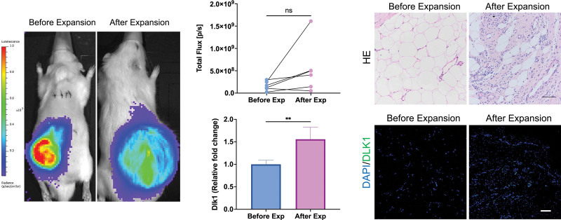
Fig. 2.
Change in transplanted adipose tissue during expansion. (Left) Luminescent images of transplanted adipose tissue in vivo. The adipose tissue was continuously observed during expansion. (Above, center) Quantified result of luminescent imaging. (Above, right) Histologic results of adipose tissue. The tissue exhibited a dense fibrotic-like appearance with a sharp decrease in adipocytes. (Below, center) mRNA expression of Dlk1 in adipose tissue before and after expansion. (Below, right) Immunohistochemical staining for Dlk1 in adipose tissue before and after expansion. Scale bar = 100 µm; **P < 0.01; ns, no significance.

