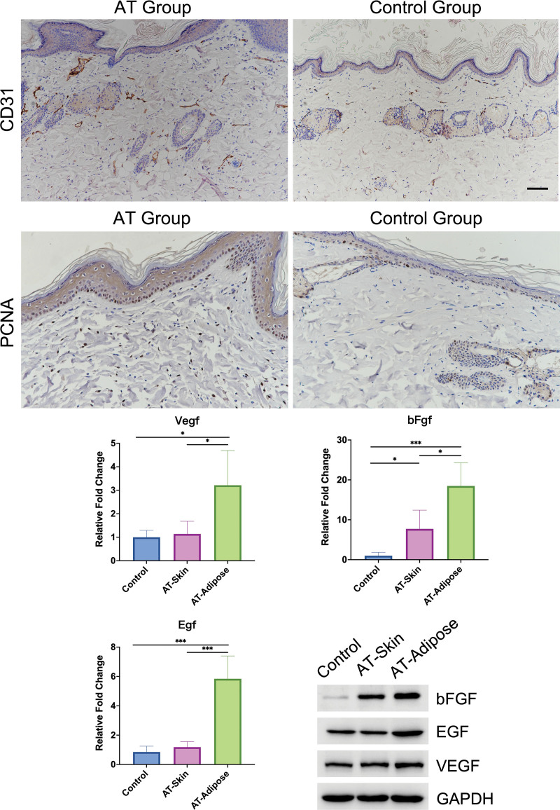
Fig. 4.
Subcutaneous adipose tissue promoted vascularization and cell proliferation in expanded skin by means of paracrine growth factors. (Above) Immunohistochemical staining for CD31 and PCNA to mark vessels and proliferating cells. (Center) mRNA expression levels of EGF, bFGF, and VEGF in skin samples from the control group and the AT group (AT-Skin), and adipose tissue from the AT group (AT-Adipose). (Below, right) Immunoblot analysis of the expression of bFGF, EGF, and VEGF in skin samples from the control group and the AT group, and adipose tissue from the AT group. Scale bar = 100 µm; *P < 0.05; ***P < 0.001.

