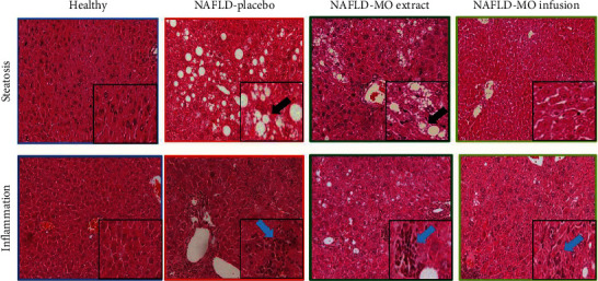
Figure 4.

Histopathological parameters in the groups. It is possible to observe that the liver tissues in the standard diet (SD) group do not show pathological changes. The NAFLD-placebo group shows a greater presence of signs of steatosis and inflammation than the liver tissues of the groups treated with NAFLD-MO. As an example, the black arrow shows an area of steatosis, while the blue arrow shows the presence of inflammatory cells. Microphotographs stained with hematoxylin and eosin at xx magnification, with the framed area presumably at ×200 magnification.
