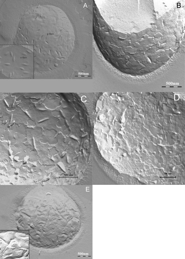
FIG. 5.
Freeze-fracture electron microscopy images. Control C. albicans cells before any treatment the showing the p face (A) and the e face (A, inset); C. albicans cells after illumination with 1.8 J · cm−2/25 μM TriP[4] showing the p face (B) and the e face (C and D); C. albicans cells after illumination with 5.4 J · cm−2/25 μM TriP[4] showing the p face (E) and after illumination with 12.6 J · cm−2/25 μM TriP[4] showing the p face (E, inset). Fluence rate, 30 mW · cm−2.

