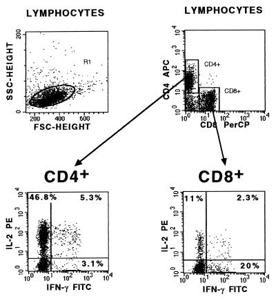
FIG. 1.
Cytokine-specific anti-human MAbs (either FITC labeled or PE conjugated) were used in combination with allophycocyanin (APC)-labeled anti-CD4 MAb and peridinin chlorophyll (PerCP)-labeled anti-CD8 MAb for intracellular staining of cells within the lymphocyte scatter gate. The numbers in each quadrant represent the percentage of gated cytokine-producing cells within the CD4+- or CD8+-cell population. The dot plots from a representative child donor show that 46.8% of gated CD4+ cells were IL-2+–IFN-γ−, 5.3% were IL-2+–IFN-γ+, and 3.1% were IL-2−–IFN-γ+. Eleven percent of CD8+ cells produced IL-2+–IFN-γ− cytokines, 2.3% produced IL-2+–IFN-γ+ cytokines, and 20% produced IL-2−–IFN-γ+ cytokines.

