FIG. 3.
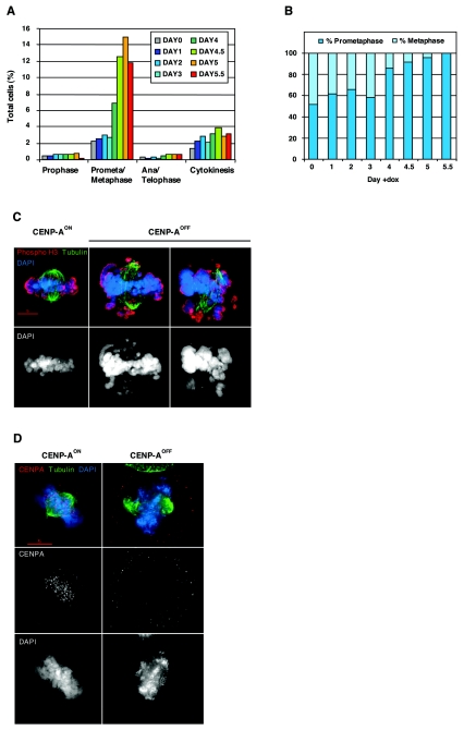
CENP-A-deficient cells are delayed in prometaphase. (A) Distribution of mitotic stages in CENP-AON (day 0) and CENP-AOFF cells during the depletion time course. Counting was performed on cells stained for tubulin, phosphohistone H3, and DNA. Five hundred to 2,000 cells, including at least 100 mitotic cells, were counted for each time point. (B) Ratio of prometaphase and metaphase cells in the CENP-AON (day 0) and CENP-AOFF populations during the depletion time course. Fifty prometaphase/metaphase cells with a bipolar spindle where both poles were localized in the same plane were counted for each time point. Equatorial chromosome alignment defined the metaphase stage. (C) CENP-AON and CENP-AOFF (doxycycline, day 4.5) were stained for tubulin (green), phosphohistone H3 (red), and DNA (blue). Metaphase cells as observed in control CENP-AON cells were rarely observed in CENP-AOFF cells. Instead, the frequency of cells with misaligned chromosomes increased. (D) CENP-AON and CENP-AOFF (doxycycline, day 4.5) were stained for tubulin (green), CENP-A (red), and DNA (blue). Kinetochore signals were no longer observed on CENP-A-deficient prometaphases.

