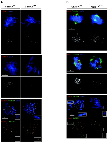
FIG. 5.
CENP-AOFF cells are depleted of the inner kinetochore proteins CENP-I, CENP-H, and CENP-C and of the Nuf2/Hec1 complex. (A) CENP-AON and CENP-AOFF metaphase spread (doxycycline, day 5) cells were stained for CENP-I (red, upper panel), CENP-H (red, middle panel), CENP-C (red, lower panel), and DNA (blue). Cells stained with CENP-C were also stained with INCENP (green, lower panel). While CENP-AON cells display strong kinetochore signals for CENP-I, CENP-H, and CENP-C, CENP-AOFF cells showed reduction of signal number and/or intensity for CENP-I and CENP-H. CENP-C signals in CENP-AOFF cells become diffuse and were no longer kinetochore localized on mitotic chromosomes. Inset panels show the inner centromere localization of INCENP and the absence of CENP-C staining for the chromosomeindicated by the box. (B) CENP-AON and CENP-AOFF prometaphase cells (doxycycline, day 5) were stained for tubulin (green), DNA (blue), and Nuf2 (red, upper panel) or Hec1 (red, middle panel). Nuf2 and Hec1 staining is strongly reduced in CENP-AOFF cells. CENP-AON and CENP-AOFF metaphase spreads (lower panel) were stained with Hec1 (Red), INCENP (green), and DNA (blue). Inset panels show that kinetochore localization of Hec1 is abolished in CENP-AOFF cells. For all immunofluorescence stainings, control slides stained with the anti-CENP-A antibody were included and no significant chromosomal signal could be detected in CENP-AOFF cells.

