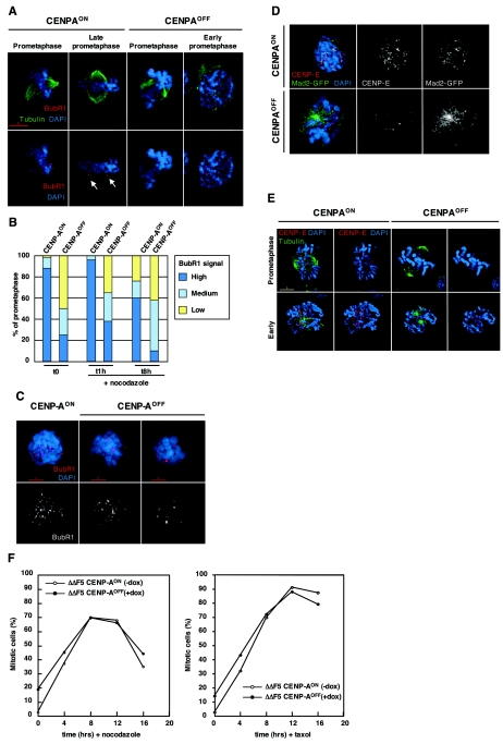
FIG. 6.
Localization of the checkpoint proteins BubR1, CENP-E, and Mad2 is affected in CENP-A-depleted cells. (A) CENP-AON and CENP-AOFF cells (doxycycline, day 4.5) were stained for tubulin (green), BubR1 (red), and DNA (blue). Unaligned chromosomes in CENP-AON prometaphase cells have strong BubR1 signals. In late prometaphase, signal intensity decreases upon chromosome alignment while out-of-the-plate chromosomes retain strong staining, as indicated by arrows. CENP-AOFF cells only retain a few kinetochores staining on misaligned chromosomes. CENP-AOFF cells still exhibit strong staining in early prometaphase. (B) Quantitation of CENP-AON and CENP-AOFF (doxycycline, day 4.5) prometaphase cells with high (many kinetochores), medium (few kinetochores), or weak (very few kinetochores) BubR1 signals. Cells were either nontreated (t = 0) or received nocodazole for 1 h or 8 h. Fifty prometaphase cells were counted in each case. (C) CENP-AON and CENP-AOFF (doxycycline, day 4.5) were treated with nocodazole (1 h) and stained for BubR1 (red) and DNA (blue). CENP-AON prometaphase cells retain a strong BubR1 staining when treated with the spindle drug, while most of CENP-AOFF cells had either few (CENP-AOFF, left panel) or very few (CENP-AOFF, right panel) kinetochores stained. (D) Nocodazole-treated (2 h) CENP-AON and CENP-AOFF cells stably expressing a Mad2-GFP construct were stained for CENP-E (red) and DNA (blue). Kinetochore localization of CENP-E and Mad2-GFP was lost in nocodazole-arrested prometaphase cells. (E) CENP-AON and CENP-AOFF cells (doxycycline, day 4.5) were stained for tubulin (green), CENP-E (red), and DNA (blue). CENP-E signals were hardly detectable in prometaphase cells, whereas some signals could be detected in early prometaphase. (F) Mitotic index of CENP-AON and CENP-AOFF (doxycycline day 4.5) during a 16-h time course of nocodazole or paclitaxel treatment. The fraction of mitotic cells was determined by flow cytometry analysis of phosphohistone H3 staining. For all immunofluorescence stainings, control slides stained with the anti-CENP-A antibody were included and no significant chromosomal signal could be detected in CENP-AOFF cells.

