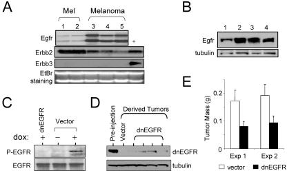
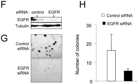
FIG. 4.
EGFR signaling is required for melanoma tumor maintenance. (A) Northern blot analysis of expression of EGFR family receptors in independent lines of cultured melanocytes (lanes 1 and 2) and independent lines of cultured melanomas from Tyr/Tet-RAS mice (lanes 3 to 5). Lane + contains positive controls for Erbb2 and Erbb3. The ethidium bromide (EtBr)-stained gel is shown as a control for equal loads in the lanes. (B) Immunoblot analysis of EGFR expression in Tyr/Tet-RAS melanomas. (C) Immunoblot analysis of endogenous levels of phospho-EGFR and total EGFR in R545 melanoma cells in vitro. Cells were transduced with dnEGFR or empty vector and grown in the presence (+) or absence (−) of doxycycline (dox). (D) Levels of ectopically expressed dnEGFR in the parental cell line (Pre-injection lane) or in tumors that emerged following injection of these cells in SCID mice (dnEGFR lanes). Lysates for the vector control are shown in the Vector lane. (E) Tumor size 14 days after subcutaneous injection with R545 melanoma cells expressing dnEGFR or empty vector. Eight tumors per time point were analyzed. Exp, experiment. (F) Western blot analysis of R545 cells transfected with control or EGFR siRNA (cell lysates were isolated 72 h posttransfection). (G) Photomicrographs showing soft-agar colony formation by siRNA-transfected R545 cells expressing H-RASV12G. (H) Tabulation of total number of colonies in the soft-agar assay.


