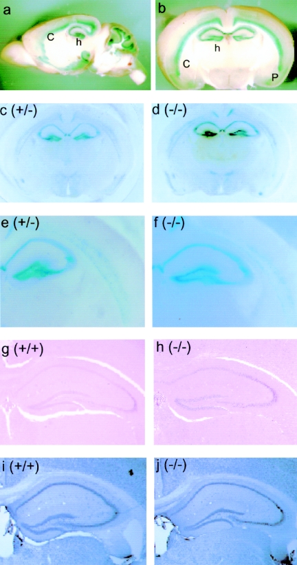
FIG. 4.
Expression of NLRR4 and hippocampal anatomy in the NLRR4−/− adult brain. NLRR4 expression was assessed by β-galactosidase staining of a sagittal section (a) and a coronal section (b) of the adult brain of 2-month-old heterozygous mice. β-Galactosidase activity was detected in the hippocampus, layers V and VI in the cortex, the piriform cortex, the inner granule layer, and Purkinje cells in the cerebellum. β-Galactosidase staining was performed in coronal sections of the NLRR4+/− and NLRR4−/− cerebrum. Similar staining patterns were observed in both NLRR4+/− and NLRR4−/− mice (c, d, e, and f). Gross hippocampal anatomy in NLRR4−/− mice was evaluated by histochemical and immunohistochemical staining. Hematoxylin-eosin staining (g and h) and immunostaining of a neuron-specific maker, NeuN (i and j), showed that there were no significant differences between the two genotypes. Abbreviations: h, hippocampus; c, cortex; p, piriform cortex.

