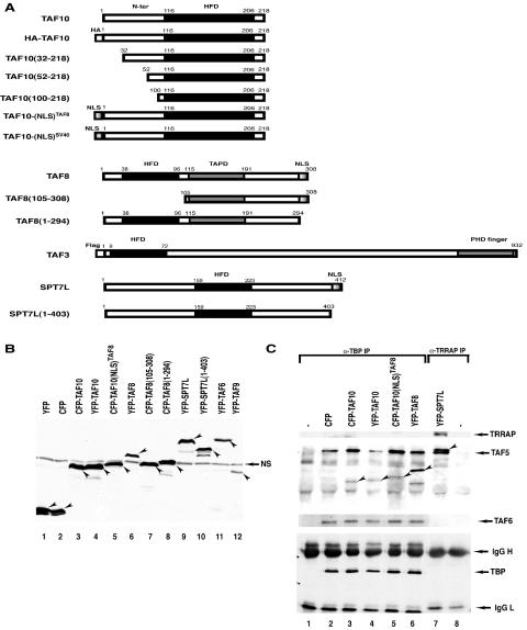
FIG. 2.
(A) Schematic representation of the TAF10-, TAF8-, TAF3-, and SPT7L-containing fusion proteins used in the HeLa cell transfection experiments. Most of the constructs were generated as CFP and/or YFP fusion proteins. The histone fold domain (HFD), the proline-rich domain of TAF8 (TAPD), the PHD finger, and the NLSs of the factors are indicated. The numbers refer to amino acid positions in each protein. N-ter, N terminus. (B) The YFP- and CFP-containing expression vectors express the correct fusion proteins in transfected HeLa cells. HeLa cells were transfected (as indicated), WCEs prepared, and 50 μg protein separated by SDS-PAGE and analyzed by Western blotting using an anti-GFP antibody. In each lane, the specifically expressed protein is labeled with an arrowhead. NS, nonspecific protein also present in nontransfected cell extracts. (C) The expressed YFP and CFP fusion transcription factors are functional since they integrate into their respective complexes. From the indicated extracts analyzed as described for panel B, either anti-TBP (lanes 1 to 6) or anti-TRRAP (lanes 7 to 8) IP was carried out, and bound proteins were analyzed as described in the legend to Fig. 1. −, antibody control without extract. The CFP or YFP fusion proteins incorporated into either a TBP- or a TRRAP-containing complex are labeled with arrowheads. Western blotting with anti-TRRAP, anti-TAF5, anti-TAF6, and anti-TBP was carried out to verify that the anti-TBP (α-TBP) and anti-TRRAP IPs worked as expected. IgG, immunoglobulin G (L, light chain; H, heavy chain).

