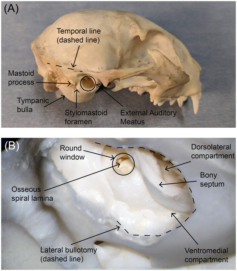
Figure 1.

Photographs of (A) the external lateral view of the feline skull and (B) the interior posteroinferior view of the tympanic bulla after bullotomy. Key anatomical markers are labeled. The area of the tympanic bulla that will be removed during an ANI procedure is traced with a black dotted line in the lower photograph. In both panels, rostral is to the right.
