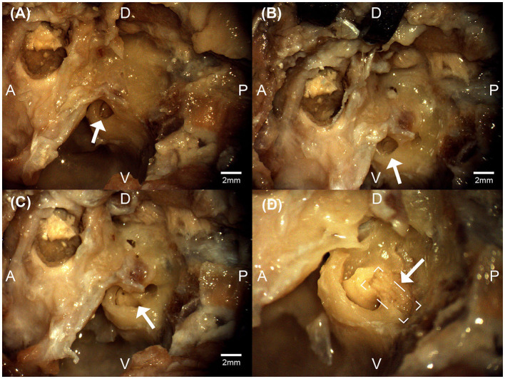
Figure 3.
Micrographs from the left bulla region of a feline cadaver exploring the transbullar translabyrinthine approach. The white arrows denote the exposed round window niche (A), the exposed cochlea (B), the exposure of the modular region (C), and the exposure of the auditory nerve (boxed) within the modiolar region (D). Anatomical directions for posterior [P], anterior [A], dorsal [D], and ventral [V] are indicated. Scale bars for (A–C) are estimated using feline cadaveric skulls' round window niche diameter. (D) is at higher magnification.

