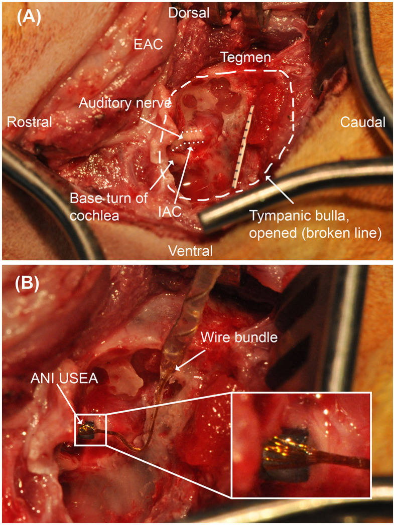
Figure 4.

Operative microscopy views of the translabyrinthine ANI procedure. (A) Surgical landmarks used to identify the auditory nerve, including the tegmen and basal turn of the cochlea are identified. Millimeter paper is placed in the field for scale. The auditory nerve target is depicted with a dotted line border. (B) 3 × 5 ANI USEA implanted into the auditory nerve. IAC, internal auditory canal. The inset box shows a close-up view of the implanted ANI USEA.
