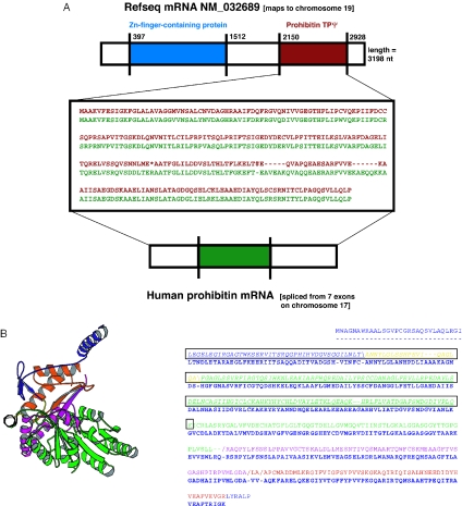
Figure 2.
Examples of TPΨgs. (A) This is a TPΨg derived from the human prohibitin gene. The prohibitin gene contains both a protein-coding region and an RNA in its 3′-UTR (45), but only the segment of the TPΨg corresponding to the protein-coding sequence is shown. In the center is an alignment of the TPΨg (in red) with prohibitin protein (in green). The graphic above it shows the position of the TPΨg (red segment) in the 3′-UTR of an mRNA that codes for a Zn-finger-containing protein (blue segment). (B) An example of a TPΨg that maps to a known globular protein domain. The TPΨg derives from the mRNA for the precursor sequence of mitochondrial 2-amino-3-ketobutyrate coenzyme A. The domain is from the closest-matching protein structure (from E.coli, PDB code 1fc4a). In the Molscript (54) picture, the protein chain trace color changes at the position of each disablement. The alignment of the E.coli domain sequence and the human TPΨg sequence is shown. The part of the sequence that maps to an EST (gi|6138420) is boxed and italicized.

