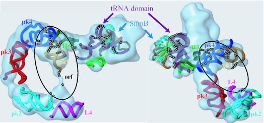
Figure 5.
Location of the reactivity changes induced when SmpB binds tmRNA onto a 3D model (two views of the model are shown). The coordinates are derived from a cryo-EM study visualizing tmRNA entry into a stalled ribosome from T.thermophilus (5). For clarity, the docking of EF-Tu into the density was omitted. SmpB (α-helices in cyan and β-barrels in light green) binds the tRNA-like domain (in dark purple) of tmRNA. H5 is in green, PK1 in orange, PK2 in light blue, PK3 in red, PK4 in dark blue and L4 in violet. The location of the reactivity changes induced when SmpB binds tmRNA are the beads. The structural alterations outside the tRNA-like domain are circled. The two figures differ by rotating the macromolecular complex along the x and y axes.

