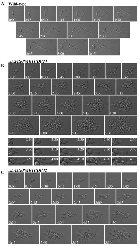
FIG. 4.
cdc24Δ/PMETCDC24 and cdc42Δ/PMETCDC42 mutants initiate a morphological response to serum but are unable to form hyphae. (A) Time lapse microscopy of wild-type cells in FCS at 37°C. DIC images were captured every 5 min over 6 h, and images every 15 min are shown over 195 min. (B) Time lapse microscopy of cdc24Δ/PMETCDC24 mutant cells in FCS at 37°C. DIC images were captured every 5 min over 6 h, and images every 15 min are shown during 330 min (top panels). Close-ups of individual cell images (bottom panels) every 10 min from 130 to 270 min are also shown. (C) Time lapse microscopy of cdc42Δ/PMETCDC42 mutant cells in FCS at 37°C. DIC images were captured every 5 min over 6 h, and images every 15 min are shown during 330 min.

