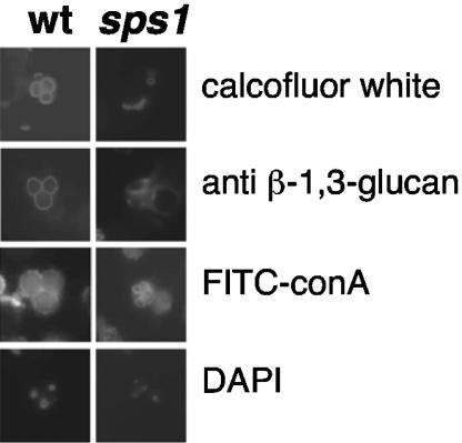
FIG. 2.
sps1 mutants do not display the full array of spore wall layers. The left panels show wild-type (wt) cells with characteristic staining patterns of calcofluor white (chitosan), anti-glucan antibodies (β-1,3-glucan), and FITC-ConA (mannan) around each individual spore. The fourth spore of the tetrad is out of the plane of focus. sps1 mutants do not display staining with calcofluor white or anti-glucan antibodies around each spore-like structure but do have distinct FITC-ConA staining.

