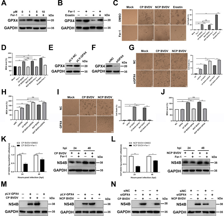
Fig 2.
Ferroptosis inhibitor inhibits BVDV-induced ferroptosis in MDBK cells. (A) Western blot analysis of GPX4 protein in Fer-1-treated and mock-treated MDBK cells at 48 h. (B) Western blot analysis of GPX4 protein in erastin-treated and mock-treated MDBK cells with or without Fer-1 at 48 hpi. (C) The percentage analysis of dead cells in mock-infected, CP BVDV (MOI = 5)-, and NCP BVDV (MOI = 10)-infected cells stained with trypan blue solution with or without Fer-1 (10 µM) at 48 hpi. Erastin (10 µM) as a positive control. Scale bar = 100 µm. (D) Detection of MDA concentrations in mock-infected, CP BVDV (MOI = 5)-, and NCP BVDV (MOI = 10)-infected cells with or without Fer-1 at 48 hpi. Erastin (10 µM) as a positive control. (E) Establishment of MDBK cell lines stably overexpressing GPX4. Western blot analysis of GPX4 proteins in overexpressing GPX4 MDBK cells and NC MDBK cells was performed. (F) Western blot analysis of GPX4 expression in MDBK cells transfected with siGPX4 or siNC for 24 h. (G) The percentage analysis of dead cells in mock-infected, CP BVDV (MOI = 5)-, and NCP BVDV (MOI = 10)-infected cells stained with trypan blue solution with or without the transfection of siGPX4 or siNC at 48 hpi. Erastin (10 µM) as a positive control. Scale bar = 100 µm. (H) Detection of MDA concentrations in mock-infected, CP BVDV (MOI = 5)-, and NCP BVDV (MOI = 10)-infected cells with or without the transfection of siGPX4 or siNC at 48 hpi. (I) The percentage analysis of dead cells in mock-infected, CP BVDV (MOI = 5)-, and NCP BVDV (MOI = 10)-infected overexpressing GPX4 MDBK cells and NC MDBK cells stained with trypan blue solution at 48 hpi. Scale bar = 100 µm. (J) Detection of MDA concentrations in mock-infected, CP BVDV (MOI = 5)-, and NCP BVDV (MOI = 10)-infected overexpressing GPX4 MDBK cells and NC MDBK cells at 48 hpi. (K) qRT-PCR and western blot analysis of viral replication and progeny in CP BVDV-infected (MOI = 5) cells with or without Fer-1 at 24 and 48 hpi, respectively. (L) qRT-PCR and western blot analysis of viral replication and progeny in NCP BVDV-infected (MOI = 10) cells with or without Fer-1 at 24 and 48 hpi, respectively. (M) Western blot analysis of NS4B proteins in overexpressing GPX4 MDBK cells and NC MDBK cells infected with CP BVDV (MOI = 5) and NCP BVDV (MOI = 10) at 48 hpi. (N) Western blot analysis of NS4B protein in mock-infected, CP BVDV (MOI = 5)-, and NCP BVDV (MOI = 10)-infected cells with or without the transfection of siGPX4 or siNC at 48 hpi. Data are given as means ± standard deviation from three independent experiments. P values were calculated using Student’s t test. An asterisk indicates a comparison with the indicated control. *, P < 0.05; **, P < 0.01; ns, not significant.

