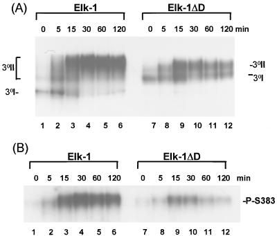
FIG. 5.
Deletion of the ERK-binding motif alters the kinetics of EGF-stimulated Elk-1 binding to the SRE. (A) COS-1 cells were transfected with 5 μg of cytomegalovirus promoter-driven expression vectors encoding either WT Elk-1 or Elk-1ΔD. Total-cell extracts were taken at the indicated times after EGF stimulation and bound to coreSRF and the c-fos SRE (SRE*). Ternary complexes containing unphosphorylated Elk-1 (3°I) and phosphorylated forms (3°II) are shown. (B) A parallel experiment was carried out with the same extracts in the presence of the anti-Phosphoplus Elk-1(Ser383) antibody. Supershifted bands representing Ser383-phosphorylated Elk-1 derivatives are indicated.

