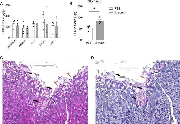
Fig 1. Ascaris suum larva hatch in and migrate through the stomach: BALB/c mice were challenged by oral gavage with 2,500 eggs of Ascaris once.
(A-B) Evan’s blue dye (EBD) was introduced intravenously to mice 1 day post infection. Recovery of EBD from (A) the intestine segments and (B) the stomach were quantified. (C) H&E and (D) PAS staining were performed on stomach sections 30 minutes post infection. Brown triangles indicate foveolar cells and black arrows indicate larval penetration across gastric mucosa. (n≥4, mean±S.E.M, *p<0.05 using two-tailed Student’s t-test. Magnification: 200×. Scale bar: 150μm. Data are shown as representative of three independent experiments).

