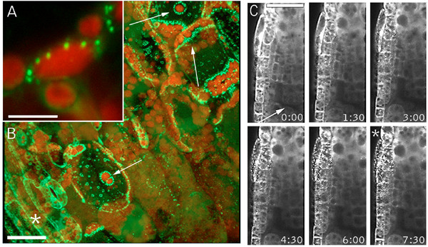
Figure 1.

Localized and transmitted wound responses of the GFP-Nit1 marker. (A, B) Aggregation in cells abutting a wound site. A 35S GFP-Nit1 leaf petiole was severed by a razor blade incision at a 45° angle to the main axis of the petiole. Three mesophyll cells exposed by the wound are indicated by the white arrows (panel B) – note the extensive redistribution of the marker in these cells to organelle-associated aggregates, which is particularly evident around chloroplasts (red fluorescence). (C) Transmitted wound response. A 35S GFP-Nit1 seedling was mounted in 0.5X MS and punctured above the root meristem (wound site marked with an arrow). Imaging was initiated after an ~45 – 60 second delay between the wound and alignment of the root tip with field of view. Aggregates become evident several cell layers removed from the wound site (marked with an asterisk). Times shown are in minutes. The image shown in A and B are 3-dimensional reconstruction of a Z-series taken 5 min after wounding. Chloroplasts are visible because of their red autofluorescence. Images in C are single optical sections. Scale bars: A 10 μm; B, C 25 μm.
