
Figure 3 |.
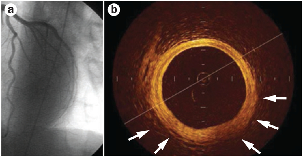
Intracoronary imaging with optical coherence tomography in a woman aged 55 years. a | Left anterior descending artery. Normal angiographic result after acute coronary syndrome. b | Optical coherence tomography shows the three-layer appearance of normal vessel wall. Intimal thickening and fibrous appearance of plaque is shown (arrows).