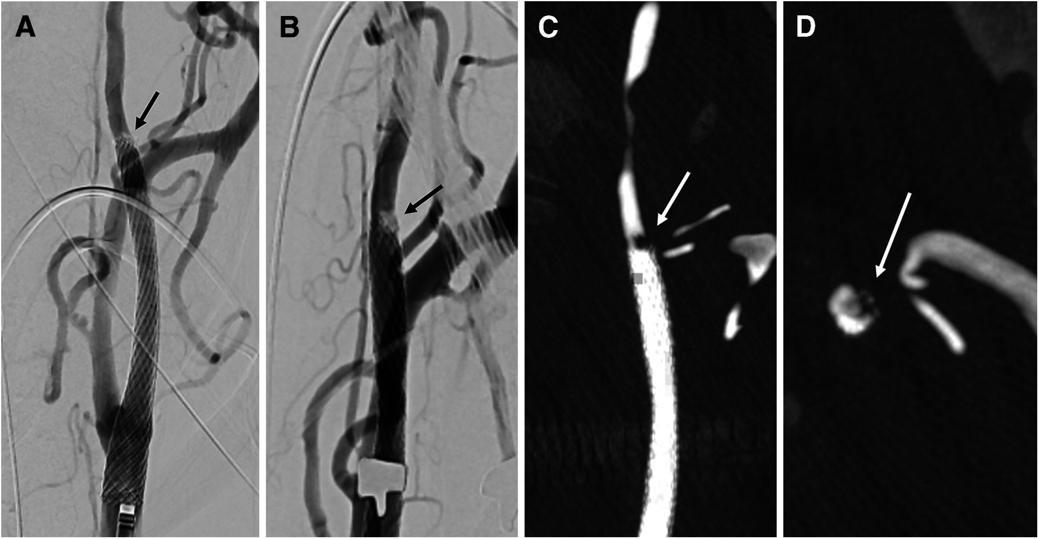
Fig. 2. (A and B) Left common carotid angiography after carotid stent deployment on anteroposterior (A) and right oblique (B) views shows a partial imaging defect at the distal end of the carotid stent in the ICA (black arrows). (C and D) 3D rotational DSA on coronal (C) and axial (D) views identifies an imaging defect (white arrows) at the same site as in panels (A) and (B). ICA: internal carotid artery.

