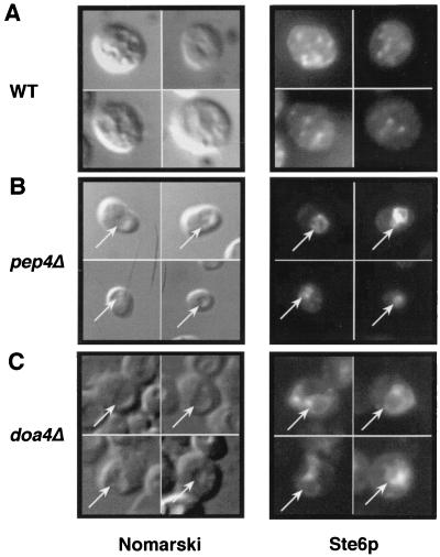
FIG. 6.
Ste6p localizes to the vacuole in the doa4Δ mutant. Ste6p localization was analyzed by immunofluorescence in a wild-type (WT) strain (SM3498; A) a pep4Δ mutant (SM2474; B), and a doa4Δ mutant (SM3501; C), all of which contain a 2μm STE6::HA plasmid (pSM693). Ste6p was detected by using antibody 12CA5. Intracellular indentations in the Nomarski image correspond to the vacuole. The arrows point to the position of the vacuole in the Nomarski images and to the position of the Ste6p signal in the middle panels.

