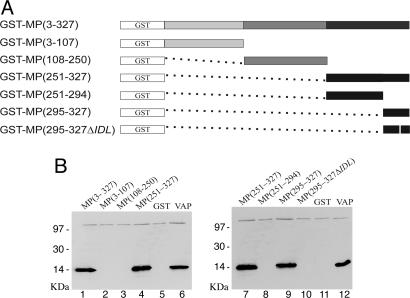
Fig. 3.
Mapping of MP domains involved in the interaction with VAP. (A)MP and MP deletion mutants are depicted by boxes of distinct colors with the construct names indicating the corresponding amino acid positions. White boxes represent GST, expressed in fusion with the proteins; black boxes represent the coiled-coil domain. Dotted lines replace deleted amino acids. The white vertical line in construct 295-327ΔIDL represents the three amino acid deletion. (B) Interaction of VAP with MP and its mutants fused to GST and with GST alone (GST, lanes 5 and 11). Proteins were separated by SDS/PAGE and detected by Western blotting with antibodies against VAP. Positions of molecular mass marker proteins are on the left of the gels. Lanes 6 and 12, VAP alone.

