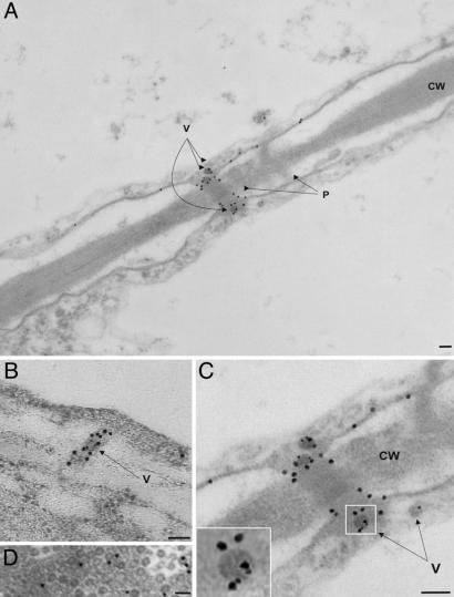
Fig. 5.
VAP and MP colocalize in modified plasmodesmata. Electron micrographs of ultrathin sections of infected tissue showing two neighboring cells. Immunogold labeling of VAP (6-nm gold particles) and MP (18-nm gold particles) shows the localization of the two proteins. (A) Two plasmodesmata (P) traversing the cell wall (CW) are indicated; one is modified and contains virus particles (V). (B) Longitudinal section of a modified plasmodesma containing CaMV particles (V) showing MP distribution. (C) Magnified image of the modified plasmodesma in A; Inset is enlarged to show MP and VAP localization on a single virion. (D) CaMV inclusion body containing virus particles. Gold particles (6-nm) label VAP. (Scale bars: 100 nm.)

