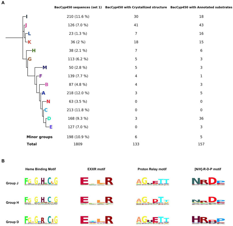
Figure 1.
Phylogenetic tree representing the major groups of BacCYPs of a sample that represents the full universe of this protein class (set 1), indicating the number of proteins in each group that have annotated structures and bound substrates or ligands (A). Configuration of groups J, H, and D, with their corresponding Hidden Markov Model (HMM) logos highlighted, reveals distinctions in the H/R amino acid from the sixth position of the Heme Binding Motif, variations in the D/E amino acid at the fourth position of the Proton Relay Motif, and differences in the N/H amino acid at the first position of the [N-H]-R-D-P motif (B).

