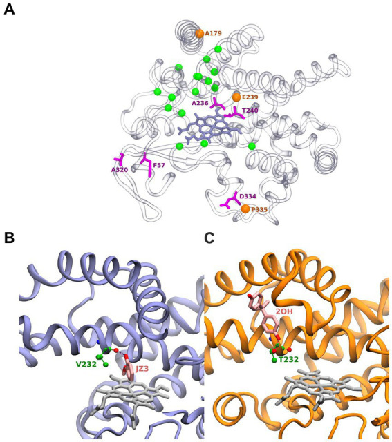
Figure 2.

Structure of BacCYPs highlighting active site residues. Residues shown as magenta sticks correspond to most conserved residues. Orange spheres correspond to positions of conserved positions where nonetheless different residues are observed establishing ligand interactions. Green spheres correspond to remaining non-conserved active site residues (A). Structure of GcoA from Amycolatopsis sp. (strain ATCC 39116 / 75iv2) bound with Guaiacol (PDBid JZ3) highlighting its interaction with the Val232 backbone (B). Cyp7863 from Streptomyces peucetius bound with Bisphenol A (PDBid 2OH) highlighting its interaction with the Thr232 side chain (C).
