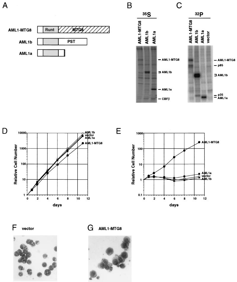
FIG. 1.
Expression of AML1-MTG8 induces G-CSF-dependent proliferation of L-G cells and inhibits their differentiation to mature neutrophils. (A) Structures of AML1-MTG8, AML1b, and AML1a. AML1-MTG8 retains the runt homology domain (Runt) of AML1 and almost all of the region of MTG8. AML1-MTG8 and AML1a lack the C-terminal transactivation domain (PST) which is present in AML1b. (B and C) Immunoprecipitation of AML1-MTG8, AML1b, and AML1a. L-G cells which expressed HA-AML1-MTG8, HA-AML1b, and HA-AML1a were labeled with [35S]methionine (B) or [32P]orthophosphate (C), and immunoprecipitations were performed with the anti-HA monoclonal antibody 12CA5. The immunoprecipitates were subjected to electrophoresis on SDS–10% polyacrylamide gels. The proteins were visualized with a BAS2000 phosphorimager (Fuji). The positions of bands of AML1a, AML1b, AML1-MTG8, CBFβ, p85, and p35 are indicated on the right. (D and E) Growth curve of the infected L-G cells in response to IL-3 (D) or G-CSF (E). The growing cells which express AML1a, AML1b or AML1-MTG8 were washed twice and cultured in the presence of 2 ng of G-CSF per ml (D) or 0.1 ng of IL-3 per ml (E). The relative numbers of viable cells are indicated. (F and G) Morphology of the cells. The vector-control (F) or AML1-MTG8-expressing cells (G) were exposed to G-CSF for 7 days and stained with May-Gruenwald’s and Giemsa’s solutions.

