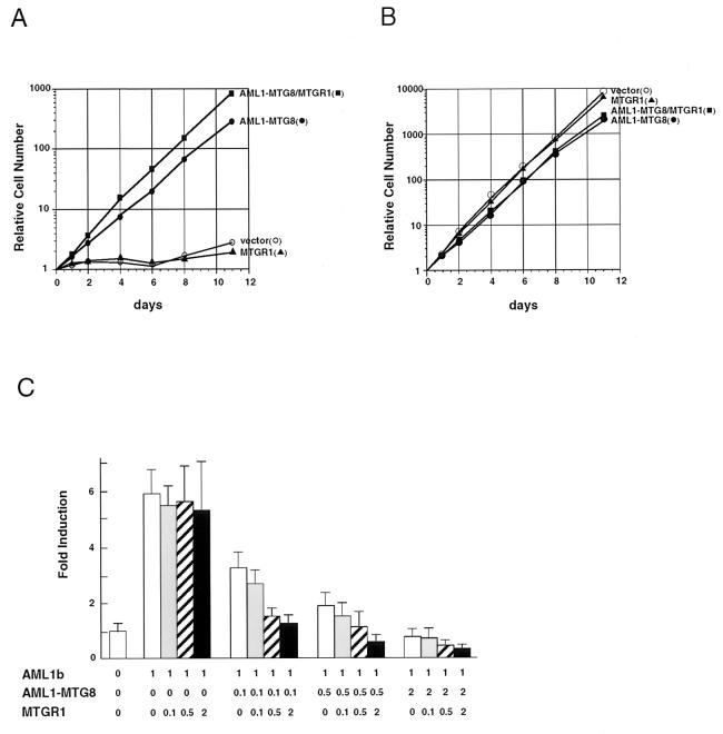
FIG. 11.
MTGR1 enhances the activities of AML1-MTG8. (A and B) Growth curve of the infected L-G cells in response to G-CSF (A) or IL-3 (B). The growing cells, which were infected with LNSX-AML1-MTG8 and/or LXSH-MTGR1, were washed twice and cultured in the presence of 2 ng of G-CSF per ml (A) or 0.1 ng of IL-3 per ml (B). Relative numbers of viable cells are indicated. (C) Repression of AML1-dependent transcriptional activation. P19 cells were cotransfected with 1.0 μg of TCRβ-TK-CAT, 1.0 μg of either pLNSX vector or pLNSX-AML1b, the indicated amounts (in micrograms) of pLNSX-AML1-MTG8 and pLNSX-MTGR1, and 0.5 μg of thymidine kinase-luciferase in a 6-cm-diameter plate. Results represent the mean relative CAT activity from three experiments which were normalized with luciferase expressed from thymidine kinase-luciferase as an internal control.

