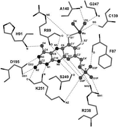
Figure 3.
The Coordination of the Moco.
Schematic representation of all protein–Moco interactions. Hydrogen bonds are drawn as dashed lines. No water-mediated hydrogen bonds between the Moco and protein were observed. The hydroxy coordination of the equatorial Mo-bound oxygen (O6) is indicated by a bold solid line and the oxo-coordination of the apical oxygen (O5) by duplicate lines. Figure was generated with LIGPLOT (Wallace et al., 1995).

