Figure 5.
The Active Site of NR-Mo.
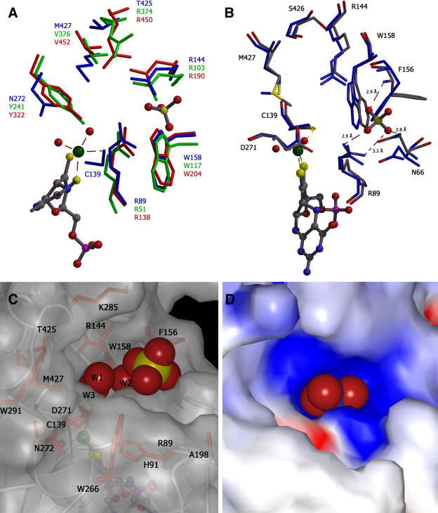
(A) Superposition of the active sites of NR-Mo2, PSO, and CSO. Residues of NR-Mo are color coded in blue, PSO in green, and CSO in red. All residues are shown in stick mode and are numbered in the corresponding color.
(B) Superposition of the active sites of NR-Mo1 (blue) and NR-Mo2 (gray). NR-Mo2 residues are colored in gray and NR-Mo1 in blue. In (A) and (B), Moco (derived from NR-Mo2) and sulfate are shown in ball-and-stick mode, and both panels were generated with MOLSCRIPT (Esnouf, 1997).
(C) and (D) Surface representation of the substrate binding site of NR-Mo2 with the bound sulfate and three active site waters (W1-3) (C) and with nitrate superimposed onto the waters (D); all are shown as a space-filling model. Surfaces are shown either transparent with highlighted and labeled active site residues or nontransparent and color coded according to the surface charge as shown in Figure 4A. The surfaces were made as described in Figure 4.

