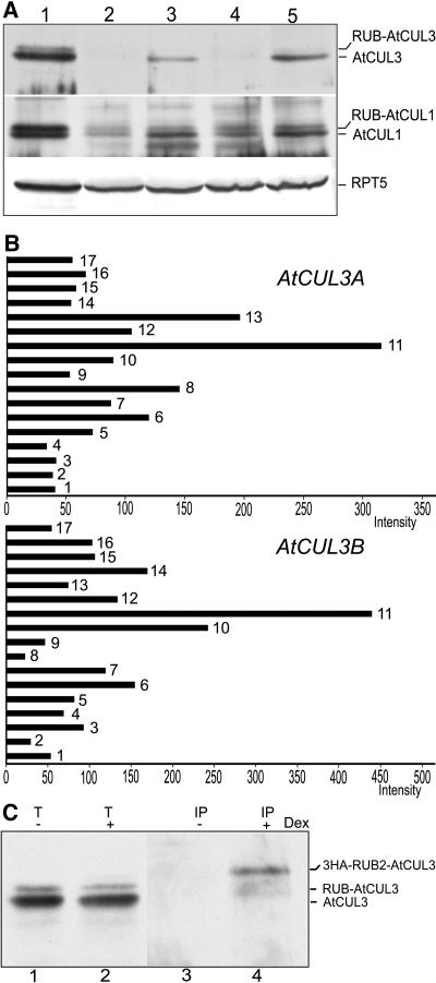
Figure 3.
Protein and mRNA Expression Patterns of the AtCUL3 Genes and Posttranslational Modification of AtCUL3 Proteins.
(A) The AtCUL3 protein is differentially expressed in Arabidopsis tissues. Total soluble protein extracts from flowers (lane 1), cauline leaves (lane 2), stems (lane 3), rosette leaves (lane 4), and light-grown seedlings (lane 5) were examined by protein gel blot analysis using anti-AtCUL3 and anti-AtCUL1 antibodies. An anti-RPT5 antibody was used as a sample loading control.
(B) Expression profiles of the two AtCUL3 genes in different Arabidopsis tissues/organs derived from a genome-profiling study using a 70-mer oligonucleotide microarray (Ma et al., 2005). The relative expression levels of each tissue/organ considered, shown with normalized intensity, are represented by black bars. Bar 1, cauline leaf; bar 2, rosette leaf; bar 3, pistil, 1 d after pollination; bar 4, pistil, 1 d before pollination; bar 5, silique, 3 d after pollination; bar 6, silique, 8 d after pollination; bar 7, stem; bar 8, sepal; bar 9, stamen; bar 10, petal; bar 11, germinated seed; bar 12, root in dark; bar 13, root in white light; bar 14, hypocotyl in dark; bar 15, hypocotyl in white light; bar 16, cotyledon in dark; bar 17, cotyledon in white light.
(C) The AtCUL3 protein is subjected to modification by RUB2 in vivo. Total protein extracts (T) from 3HA-RUB2 transgenic seedlings with (+) or without (−) dexamethasone (DEX) induction were subjected to immunoprecipitation (IP) with anti-HA antibodies. Protein gel blot analysis was subsequently performed using anti-AtCUL3 antibodies.

