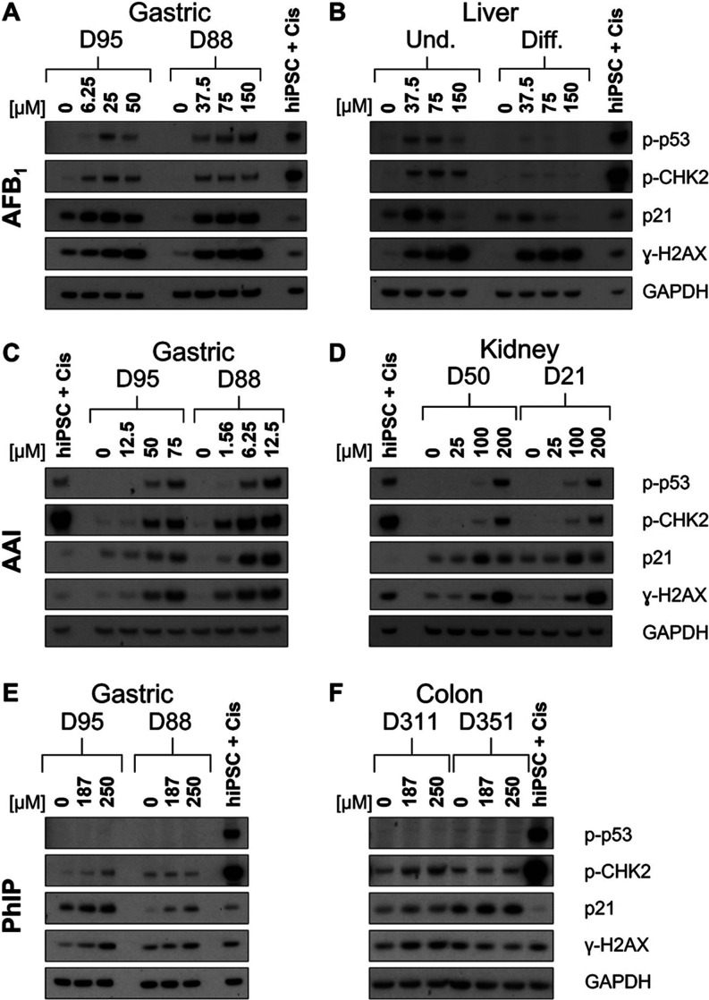
Figure 3.
DDR in normal human tissue organoids treated with AFB1, AAI, and PhIP. Organoids from gastric (D95 and D88; A, C, and E), liver (D4 undifferentiated and differentiated; B), kidney (D50 and D21; D), and colon (D351 and D311; F) tissues were treated with the indicated concentrations of AFB1 (A, B), AAI (C, D), and PhIP (E, F) for 48 h, and lysates were analyzed by Western blotting. Various DDR proteins (p-p53, p-CHK2, p21, and γ-H2AX) were detected, and GAPDH was used as a loading control. iPSC + Cis (hiPSC treated with 3.125 μM cisplatin) was used as the positive control. Representative blots are shown (n = 2).

