Fig. 2.
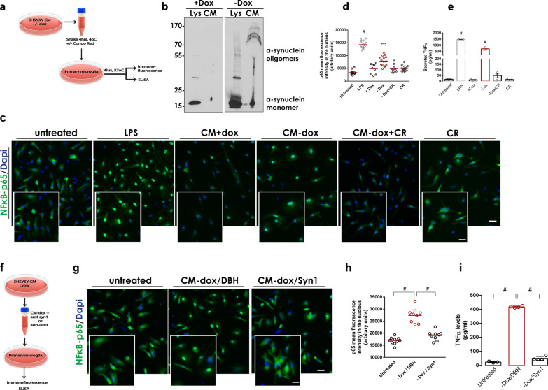
Cell-produced α-synuclein oligomers, but not monomers, can activate primary microglia. a, f Schematic representations of the experiments with primary microglia treated with CM from SH-SY5Y α-synuclein expressing cells. b Immunoblot showing the presence of cell-secreted (CM) and intracellular (Lys) α-synuclein conformers in the absence of doxycycline (− Dox). c, g Representative images of primary microglia immunostained with a specific antibody against NF-κB. DAPI was used as a marker for nuclei. Magnification of the boxed area is shown for each merged image. Scale bar: 20 μm (50 μm for the magnified images). d, h Quantification of mean fluorescence intensity of nuclear NF-κB (N ≥ 9 for all treatments). e, i Measurement of secreted TNFα from primary microglia upon indicated treatments (n = 3 biological replicates). In d, e, h, and i, statistics was performed by one-way ANOVA followed by Tukey’s multiple comparisons test. In d, #P < 0.0001 for untreated vs LPS, ***P = 0.0001 for + Dox versus − Dox. In e, #P < 0.0001 for untreated versus LPS and + Dox versus − Dox. In h and i, #P < 0.0001

