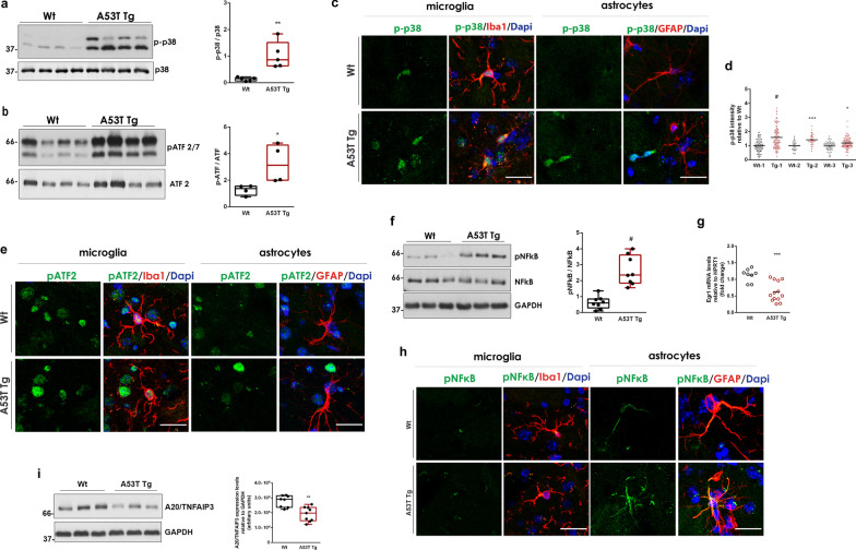
Fig. 4.
The p38MAPK and the NF-κB pathways are activated in A53T Tg striatum. a Immunoblotting analysis and quantification of phospho-p38MAPK levels relative to total p38MAPK expression (n = 5 mice per genotype, #P < 0.0001). b Immunoblotting analysis and quantification of phospho-ATF2/7 levels relative to total ATF2 expression (n = 4 mice per genotype, *P = 0.0378). c, e, h Representative magnified confocal images of striatal sections co-stained with antibodies against phospho-p38 (c), phospho-ATF2/7 (e) and phospho-NF-κB (h) and Iba1 or GFAP. DAPI (blue) was used for nuclei staining. Scale bar: 20 μm. d Measurement of phospho-p38 mean fluorescence intensity in Iba1+ cell surfaces (n ≥ 35 cells per mouse for three independent Wt-A53T Tg animal pairs, #P < 0.0001, ***P = 0.0004, *P = 0.0239). f Representative immunoblot and quantification of phospho-NF-κB relative to total NF-κB in striatal homogenates (n = 8 mice per genotype, #P < 0.0001). g qPCR measurement of Egr1 expression (n ≥ 13 per genotype, ***P = 0.0004). (i) Immunoblotting detection and quantification of A20/TNFAIP3 (n ≥ 8 per genotype, **P = 0.0011). GAPDH was used as a loading control. All statistics by unpaired Student’s t test

