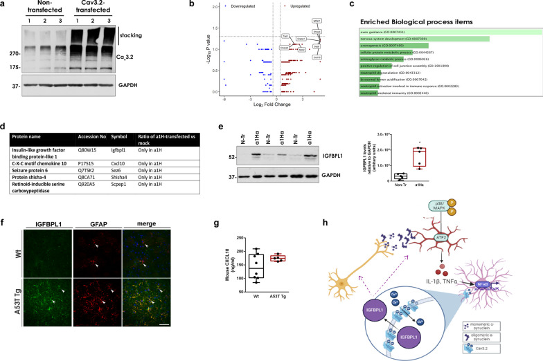
Fig. 7.
The induction of Cav3.2 VGCCs alters astrocyte secretome promoting the release of the neuroprotective protein IGFBPL1. a Representative immunoblot of mock- and a1H-transfected primary quiescent astrocytes using an antibody against Cav3.2. b Volcano plot showing the up- and down-regulated proteins identified by SignalP as secreted proteins identified by the LC–MS/MS analysis from the CM of mock- and a1H-transfected astrocytes (n = 3 independent replicates). c List of the top ten pathways as classified by Enrich-r. d List of the top five secreted proteins identified in the CM of astrocytes after a1H overexpression. e Immunoblot from mock and a1H-transfected astrocytes using an antibody against IGFBPL1 and densitometric quantification (n = 5 per condition, *P = 0.0203). f Representative confocal images of striatal sections co-stained with antibodies against IGFBPL1 and GFAP. DAPI (blue) was used to stain nuclei. Scale bar: 50 μm. g Quantification of mouse CXCL-10 levels in striatal homogenates (n ≥ 5, P = 0.1864). h Proposed mechanism through which α-synuclein oligomers activate microglia and astrocytes and induce Cav3.2 channels that mediate IGFBPL1 secretion. GAPDH was used as a loading control. In e, g, statistics were performed by Unpaired Student’s t test

