Fig. 1.
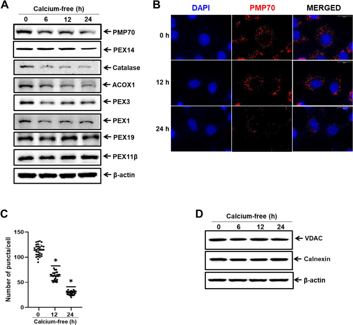
Calcium deficiency induces peroxisomal protein degradation in a time-dependent manner. A Immunoblot analysis of AML12 cells in calcium-deficient medium for indicated durations. Whole cell lysates were reacted with anti-catalase, anti-Pex14, anti-PMP70, anti-Acox1, anti-Pex3, anti-Pex1, anti-Pex19, anti-Pex11β, and anti-β-actin for protein expression analyses. B AML12 cells in calcium-deficient medium for indicated durations were fixed and immunostained with anti-PMP70 (red), shown as representative fluorescence image. Scale bar represents 25 µm. C Quantification of PMP70 puncta represents the number of peroxisomes per cell. Data are expressed as means ± S.D. (n = 3, independent experiments, 30 cells were analyzed in each experiment), * p < 0.05. D Immunoblot analysis of AML12 cells in calcium-deficient medium for indicated durations. Whole cell lysates were reacted with anti-VDAC, anti-Calnexin, and anti- β-actin

