
Fig. 5.
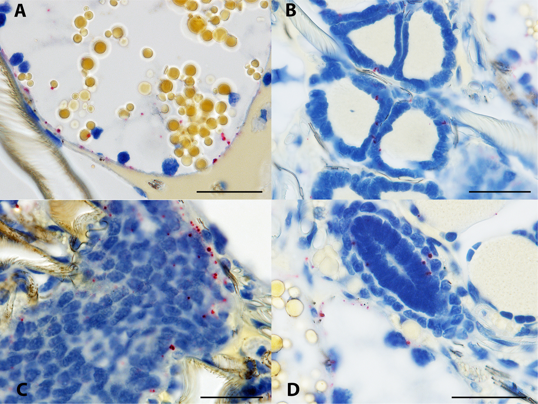
Borrelia miyamotoi-infected female Ixodes scapularis subjected to in situ hybridization assay. (A) spirochetes are apparent near the basal lamina of a distal diverticulum of the midgut, recognizable by the many straw-colored inclusion bodies evident within the digestive cells. (B) spirochetes are present within the densely packed epithelial cells of a Malpighian tubule. (C) spirochetes are present in the ovarian tissue of an unfed female. (D) spirochetes are apparent within and in the vicinity of an oviduct, shown as the ovaloid cluster of nuclei center image. 16S rDNA probe hybridization to spirochetes is indicated by red staining, blue indicates nuclear staining. Scale bars = 30 μm.