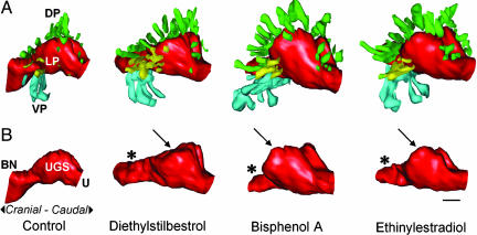
Fig. 2.
3D serial section reconstruction of the UGS from GD-19 mice exposed to low doses of estrogenic chemicals, DES, bisphenol A, and ethinylestradiol, during fetal development. The UGS depicted for each treatment was closest to the group mean for prostate duct number and size. All images are viewed from a left-lateral perspective. (A) Shown are the differences in patterns of prostate ductal development after fetal exposure to these chemicals, compared with oil-treated controls. There is a significant increase in the total number of ducts in estrogen-treated animals and a corresponding increase in overall prostate volume. (B) Shown is the marked alteration in the shape of the urethra (U, red) in the region of the bladder neck (BN), which is markedly constricted (*) inthe mice exposed to the estrogenic chemicals, compared with controls. In addition, the region of the UGS (the prostatic sulcus or colliculus, arrow) associated with the development of the dorsal (DP, green) and lateral (LP, yellow) prostate ducts is enlarged, particularly by bisphenol A, compared with controls. Ventral prostate (VP, light blue). (Scale bar, 100 μm.)

