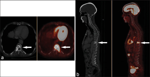
Figure 2.

(a) Axial images and (b) sagittal images in the CT bone window and fused PET/CT in white arrows. The whole-body 18F-FDG PET/CT scan reveals a metabolically active (SUVmax 11.2) expansile-enhancing soft tissue osteolytic lesion with dense interspersed sclerosis in the body of the D10 vertebra contiguously involving its posterior elements with cortical erosions and solid periosteal reactions. Mild erosions of the adjoining right 10th rib were also noted
