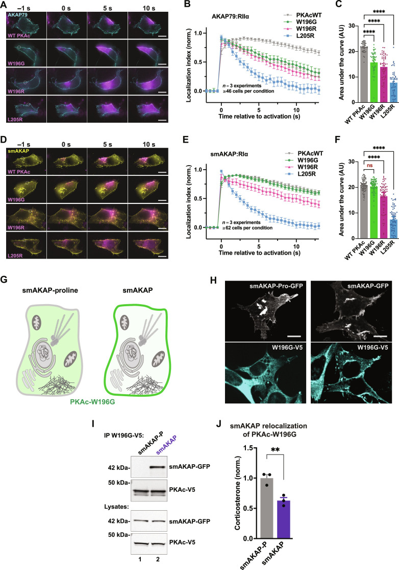
Fig. 7. Sequestrating PKAcW196G corrects cortisol overproduction.
(A to F) Type II (A) and type I (D) photoactivation time courses in H295R cells. PKAc variants tagged with photoactivatable mCherry were expressed along with either AKAP79-YFP and RIIα-iRFP [(A) to (C)] or small membrane-bound AKAP (smAKAP)–green fluorescent protein (GFP) and RIα-iRFP [(D) to (F)]. Plotting PKAc localization after photoactivation [(B) and (E)] and area under the curve [(C) and (F)] demonstrates differences among mutants. Localization index = [(intensity of activated region − background intensity)/(intensity of cytosolic region 6 to 8 μm distal − background intensity)]. Scale bars, 10 μm; means ± SE; n = 3 replicates with a total of at least 46 [(A) to (C)] and 62 [(D) to (F)] cells per condition; ****P ≤ 0.0001, one-way ANOVA with Dunnett’s correction. ns, not significant. (G) Cartoon depiction of experimental design for the PKA anchoring defective smAKAP-proline mutant (left) and smAKAP (right) sequestration experiments. Green demarks expected localization of PKAc-W196G. (H) Fluorescent images of ATC7L cells stably expressing PKAc-W196G-V5 (cyan) along with GFP-tagged constructs of either smAKAP-proline (white) or WT smAKAP (white). Scale bars, 10 μm. See also fig. S4. (I) Immunoprecipitation of PKAc-W196G from stable W196G/smAKAP-proline (lane 1) or W196G/smAKAP (lane 2) ATC7L adrenal cells. Representative of three biological replicates. (J) Corticosterone measurements from stable ATC7L cells coexpressing PKAcW196G with either smAKAP-proline (gray) or smAKAP (purple). Means ± SE; n = 3; **P ≤ 0.01, Student’s t test.

