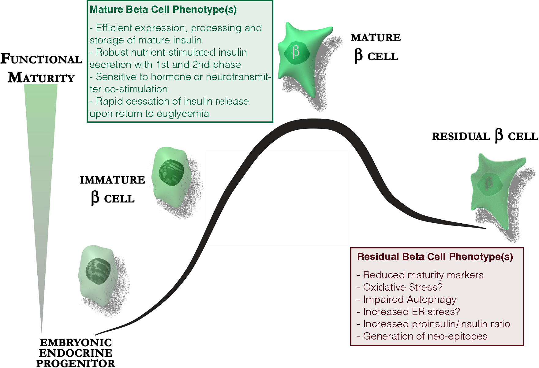
Figure 1: Residual beta-cells in type 1 diabetes are dysfunctional.

The majority of beta-cells in long-standing type 1 diabetes are lost. Residual beta-cells express lower levels of maturity markers, experience increased oxidative stress and have impaired autophagy. They demonstrate increased ER stress as a consequence of the increased demand for proinsulin biosynthesis placed upon a dwindling number of beta-cells. This is associated with increased release of incompletely processed proinsulin and could lead to the generation of neo-epitopes in the beta-cell specific autoimmune response.
