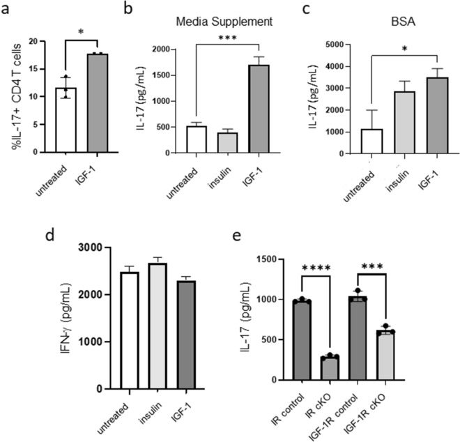
Figure 2.
IGF-1 treatment increases IL-17 production by CD4+ T cells. (a) CD4+ T cells were activated for 48 h on anti-CD3/CD28 coated plates in serum free conditions in the presence or absence of IGF-1 for the last 24 h of activation, after which IL-17 expression was analyzed by flow cytometry. (b, c) CD4+ T cells were activated for 48 h on anti-CD3/CD28 coated plates in media supplemented with either Insulin Free Media Supplement (b) or 0.35% BSA (c) in the presence or absence of insulin or IGF-1 for the last 24 h, after which IL-17 production was measured by ELISA. (d) CD4+ T cells were activated for 48 h on anti-CD3/CD28 coated plates in media supplemented with Insulin Free Media Supplement in the presence or absence of insulin or IGF-1 for the last 24 h, after which IFN-γ production was measured by ELISA. (e) Splenic CD4+ T cells from IR cKO or IGF-1R cKO and littermate control mice were activated for 48 h in full serum conditions, after which IL-17 production was measured by ELISA. Data representative of at least 2 independent experiments; n = 3 mice per experiment. Data analyzed using student’s t-test (*p < 0.05; ***p < 0.001 ****p < 0.0001).

