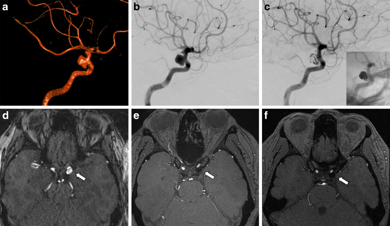
Fig. 2.
a 3D-angiography showing a saccular multilobulated aneurysm of the left internal carotid artery. b Subtracted digital subtraction angiography (DSA) before endovascular treatment. c Subtracted and unsubtracted DSA after coiling and FRED implantation showing residual aneurysm filling. d Magnetic resonance imaging (MRI) time of flight angiography (TOF) angiography before treatment with an arrow pointing to the aneurysms. e 1-year follow-up MRI TOF-angiography showing punctate residual perfusion of the aneurysm neck. f 6-year follow-up MRI TOF-angiography showing stable complete occlusion of the aneurysm

