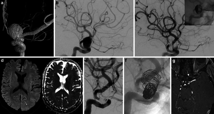
Fig. 3.
a 3D-angiography showing a large (maximum diameter 14 mm) saccular aneurysm in the paraophthalmic left ICA. b Subtracted DSA before endovascular treatment. c Subtracted and unsubtracted DSA after coiling and FRED implantation showing stasis in the aneurysm. d MRI scan after 24.9 months from the index procedure due to several episodes of amaurosis fugax while being under ASS monotherapy: DWI and ADC maps showing a punctate diffusion restriction (arrows) restriction in the left occipital lobe. e , f Subtracted and unsubtracted DSA of the same day showing the patency of the FD and adjacent vessels, very mild intimal hyperplasia, along with stable complete occlusion of the aneurysm. g MRI TOF sequence showing complete aneurysm occlusion at the 5‑year follow-up

