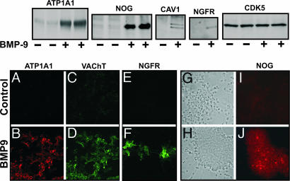
Fig. 3.
Analysis of BMP9-induced proteins. (Upper) Western blots of selected proteins from control and BMP9-treated neuronal cultures. Septal cultures from E14 mice were treated for 3 days with BMP9 (10 ng/ml) or vehicle. Cells were harvested and processed for SDS/PAGE as described in Materials and Methods. (Lower) Immunocytochemistry (A–J) of cultures are treated as in Upper.(A–D) Double immunofluorescence staining with anti-Na+/K+ ATPase α-1 and anti-VACHT antibodies, done in parallel with negative and positive controls. (E and F) Immunofluorescence staining with anti-NGFR antibody. Phase-contrast (G and H) and immunofluorescence (I and J) staining with anti-Noggin antibody of the same field. All pictures were obtained with a ×20 objective.

