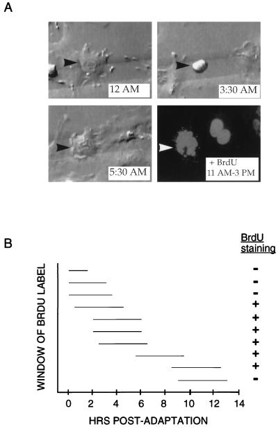
FIG. 3.
Timing of S phase entry in p53−/− MEFs treated with nocodazole. (A) Time-lapse videomicroscopy and subsequent immunofluorescence of representative p53−/− MEF that arrested at mitosis in the presence of nocodazole. The cell initiated mitotic arrest starting shortly after 12 AM and then adapted at 5:30 AM (top panels and lower left panel). At 11 AM, the BrdU label was added to the media; the cell was then recorded for an additional 4 h and fixed. Immunofluorescence was performed to detect BrdU incorporation (lower right panel). As indicated by arrowheads, the same cell was identified during video recording and immunofluorescence by its position on a gridded coverslip. (B) Measurement of time of S phase entry relative to time of adaptation from mitotic arrest. Ten p53−/− MEFs on gridded coverslips were treated with nocodazole, monitored by time-lapse videomicroscopy, and pulsed with BrdU for 4 h at various times following adaptation. For each cell, the horizontal line indicates the period during which BrdU was present (measured in hours) relative to the time elapsed since the cell underwent adaptation. The + or − indicates whether the cell stained positive for BrdU incorporation by immunofluorescence.

