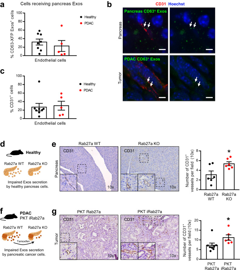
Fig. 3. Pancreas exosomes mediate local communication and constrain angiogenesis.
a Dot plot representing the percentage of endothelial cells (CD31+) that received pancreas-derived CD63+ Exos in health (Panc-CD63-mCherry, n = 8) and in PDAC (KPF CD63-mCherry n = 2 and KPC-ExoBow n = 3) analyzed by flow cytometry. b Representative confocal microscopy images of pancreas-derived Exos (green) accumulation in endothelial cells (CD31+ in red) in healthy pancreas of Panc-CD63-mCherry mice (upper panel) or tumors of KPF CD63-mCherry mice. Nuclei were counterstained with hoechst (blue). Scale bar 5 μm. c Dot plot representing the percentage of endothelial cells (CD31+) in the pancreas microenvironment in health (Panc-CD63-mCherry, n = 8) and in PDAC (KPF CD63-mCherry n = 2 and KPC-ExoBow n = 3) analyzed by flow cytometry. d Schematic representation of the healthy Rab27a KO GEMM in which pancreas cells have impaired secretion of exosomes. e Representative CD31 IHC images (10 x, left) and respective quantification (right) in the pancreas of wild-type (Rab27aWT, n = 6) and Pdx1 Rab27aFrt/Frt (Rab27aKO, n = 6) mice. Two-tailed unpaired t-test, p = 0.0261. f Schematic representation of the PDAC Rab27a KO GEMM in which pancreas cells have impaired secretion of exosomes upon tamoxifen administration. The PKT Rab27a model is the control group which lacks the R26LSL-Flpo-ERT2 allele, hence upon tamoxifen administration expresses Rab27a and has proficient exosomes secretion. g Representative CD31 IHC images (10 x, left) and respective quantification (right) in the pancreas of control PKT Rab27a (n = 8) and PKT iRab27a (n = 6) mice. Two-tailed unpaired t-test, p = 0.0485. Data are Mean ± SEM. Source data are provided as a Source Data file. Schemes created with BioRender.com.

